
Heat Diffusion Imaging evolves as new study shows AI can classify specific canine tumour types
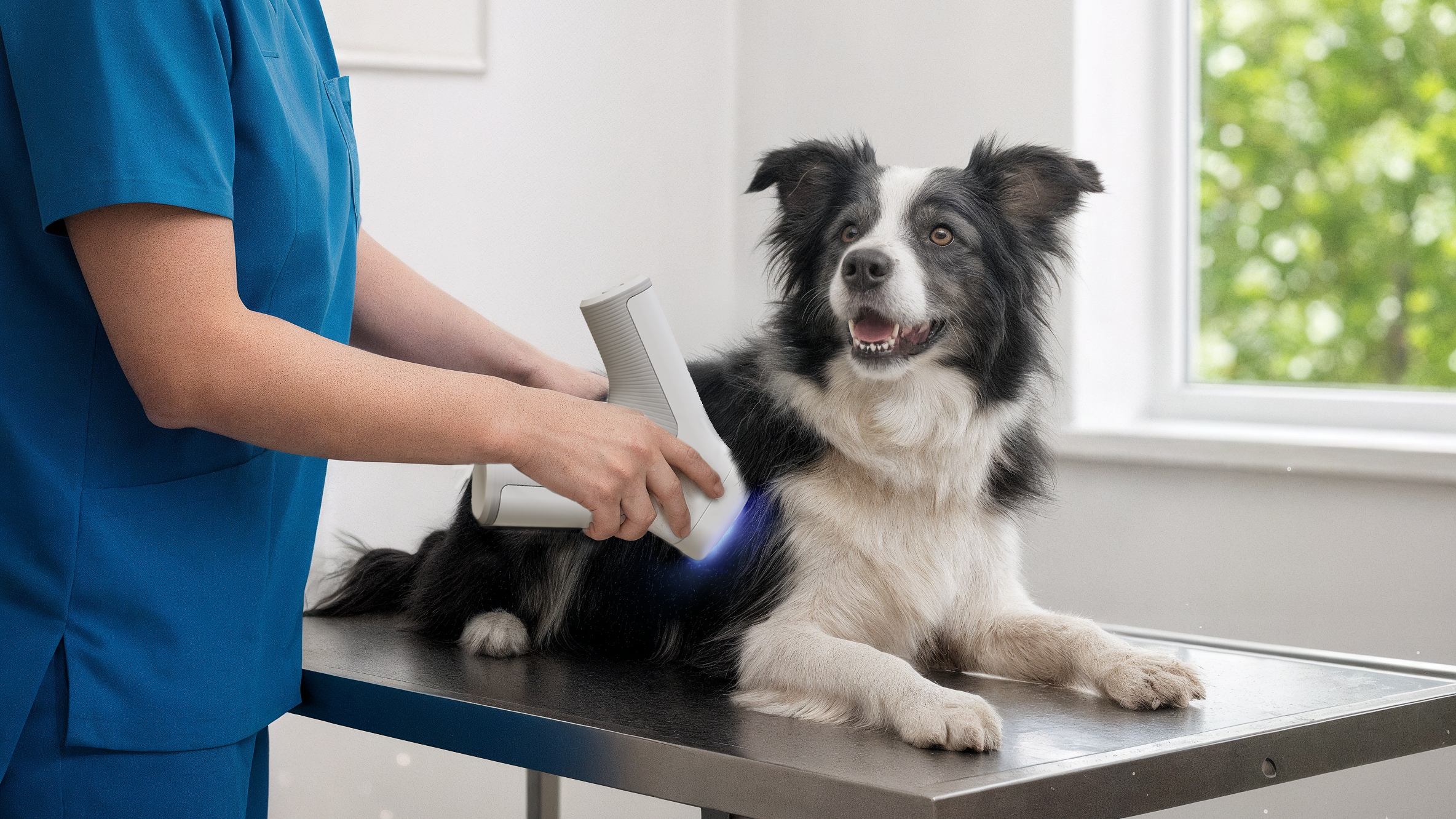
Peer-Reviewed study confirms Heat Diffusion Imaging used by non-invasive AI screening model screens canine cancer with 98% NPV at an assumed 15% malignancy rate as over 500 practices adopt the scanner
New peer-reviewed research shows HT VET’s Heat Diffusion Imaging (HDI) technology moving beyond benign vs malignant screening, with new AI classifiers designed to identify specific tumour types. The study, published in Veterinary Sciences on September 9, 2026, “Tumor-Specific Classification of Canine Cutaneous and Subcutaneous Tumors Using Heat-Diffusion Imaging and Artificial Intelligence,” led by HT VET Chief Veterinary Officer Dr. Gillian Dank, evaluated 1,010 masses in 669 dogs. It shows the HDI technology used in the non-invasive scanner, Vista iQ, rules out malignancy in canine lumps and bumps with 92% sensitivity and a 98% negative predictive value (NPV) at an assumed 15% malignancy rate.
Building on this screening role, the study tested whether VISTA iQ could provide additional information about masses already classified as highly likely to be benign or malignant. Two new tumour-specific classifiers target lipomas and mast cell tumours (MCTs), the most common benign and malignant skin tumours in dogs.
“With the majority of canine lumps and bumps currently left untested, veterinary medicine has lacked an immediate and non-invasive standard for early screening,” said Shani Toledano, Co-founder and CEO of HT VET. “This research demonstrates not only the strength of our technology in helping veterinary teams rule out malignancy, but the potential for AI to provide increasingly sophisticated insights into the masses they encounter every day.”
In the held-out test set, all 38 masses flagged by the lipoma classifier were confirmed as lipomas (86.4% sensitivity, 100% specificity).
The MCT classifier was designed to prioritise specificity over sensitivity. It flags fewer masses, but those it flags are very likely to be malignant and most are mast cell tumours (14 of 16 in the test set). An MCT flag can prompt antihistamine cover before aspiration and early staging, and gives owners a clear reason to proceed. Importantly, a negative MCT result does not rule out a mast cell tumour, and these masses should still be investigated as the primary classifier indicates.
Toledano added, “As our technology learns and evolves, our aim is to give veterinary teams more information at the earliest stage of the diagnostic pathway, helping them decide which masses can be monitored and which need further investigation.”
The findings represent an important preliminary validation milestone for tumour-specific classification and demonstrate the potential for dynamic thermal imaging combined with AI to provide increasingly refined information at the point of care.
The publication comes as adoption grows. VISTA iQ is now used in more than 600 veterinary practices and organisations worldwide, with more than 40,000 scans performed to date.
Customer data points to increased diagnostic activity. In HT VET’s April 2026 survey 77 practices using HT VET’s technology, 91% reported testing more masses and 74% reported diagnosing more malignancies. This shows a shift in mindset with more lumps being tested and more malignancies subsequently being found. HT VET look to independent validation being the next step.
The full peer-reviewed study is available here: https://www.mdpi.com/2306-7381/13/9/932